Parts Of Foot Bones (Anatomy, Skeleton, Pictures)

Last updated on July 26th, 2018 at 04:22 pm
Question: What are the parts of foot bones?
Learning your foot anatomy is important especially to know which bone is currently causing foot pain.
Also, for students and health professionals, it is critical to understand the foot anatomy which basically improves your knowledge of bone position, ligament attachment, and the way tendons run on the foot bones.
The foot is made up of 26 bones, 33 joints, and over 100 ligaments, tendon, and muscle
What you will learn in the article
- The parts of the foot bones
- The different bones on each section of the foot
Parts of foot bones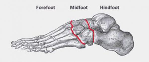
The foot is located after the long shin bones and it starts from the back of your ankle to your toes. Anatomically, the foot is divided into 3 sections. The hindfoot, midfoot, and the forefoot.
The hindfoot is the posterior part of the foot. “Hind” means posterior, so, it basically the backward part of the foot. The hindfoot consists of bone from the leg and the ankle joint. The lower ends of the tibia and fibula, the calcaneus, and talus bones are located at the hindfoot
In a similar way, “mid” means the middle of the foot. The midfoot is just in front of the hindfoot. This part of the foot consists of bones that make the arch of the foot. The foot arch is a small elevation at the middle of the foot and its critical for supporting your body’s weight
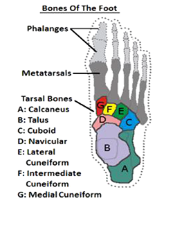
The midfoot bones are the navicular, cuboid, and 3 cuneiform bones
The forefoot is the furthermost part of the foot and consist of metatarsal bones and phalanges that make up your toes.
The foot bones are generally grouped into tarsal, metatarsal and phalanges. So, to simplify, the hindfoot and midfoot consist of 7 tarsal bones (calcaneus, talus, navicular, cuboid, and 3 cuneiforms) while the forefoot consist of 5 metatarsal bones and 14 phalanges.
Hindfoot Bones Anatomy
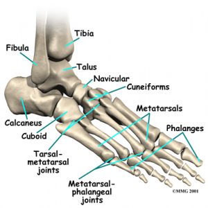
foot skeleton picture
Like already mentioned, the hindfoot is the posterior part of the foot. Its made up of 4 bones; The calcaneus, talus, fibula, and tibia bones.
1. The tibia bone
The tibia is one of the 2 bones that make up the leg. It extends from your knee joint upwards to the ankle joint downwards. The tibia bone makes 4 joints in the body
- The knee joint
- The ankle joint
- The superior tibiofibular joint (joint near the knee that holds the tibia and fibula together)
- The inferior tibiofibular joint (the lower joint at the ankle that holds the tibia and fibula together)
The tibia is the second largest bone in the body and is firmly located in the inner middle part of the leg. At the foot, it articulates with the talus and fibula to form the ankle joint.
At the lower end, the tibia bone expands, forming a prominence on the inside of the foot. This is called the medial malleolus and is critical for attachment and passage of tendons.
2. The fibula bone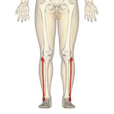
The fibula is the second smaller shin bone and extends from the knee down to the ankles. Like the tibia, there are 2 fibula bones in your body. The right and left fibula bones.
However, unlike the tibia bones, the fibula bone is not involved with weight bearing though helps in the stability of the ankle joint.
At the knee, the fibula bone articulates with the tibia bone (superior tibiofibular), then extends downward to ankle and enters the hindfoot.
Most importantly, the fibula bone forms a lateral prominence on the outside of the foot (lateral malleolus) which is also important to ensure ankle stability
3. The calcaneus (heel bone)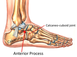
The calcaneus is the largest foot bone and is located at the back of the hindfoot. It is important for the attachment of the Achilles tendon which attaches to the posterior part of the heel bone.
At the lower part of the heel bone is the plantar ligament that supports your body’s weight. At the upper part of the heel bone, it articulates with the talus. In front, it articulates with the cuboid bone (calcaneocuboid joint).
4. The Talus bone
The talus bone is the second largest bone in the entire foot, and unlike the rest bones, there is no attachment of muscles. It forms the lower part of the ankle (formed collectively by the tibia, fibular, and talus bones).
Primarily, it functions to transmit your body’s weight to the foot through the talocalcaneonavicular joint. This joint is formed by the talus, calcaneus, and the navicular bones.
The talus bone may also be fractured. If it does, healing is difficult due to the decreased blood supply to that part of the foot.
parts of foot midfoot
The midfoot Bones Anatomy
The midfoot is made up of 5 tarsal bones. Together with the metatarsal bones (proximal bones in the forefoot), they form the arch of the foot.
The midfoot bones are the navicular, cuboid and cuneiform bones
1. Navicular bone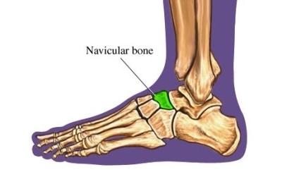
The navicular bone is one of the midfoot bones. It is located on the inside of the foot and rarely gets fractured.
Attachment: Posteriorly the navicular bone is attached to the talus bone. In front, it articulates with the cuneiform bones while on the outer surface it articulates with the cuboid bone.
The navicular bone may get fractured from repetitive use or direct injury. If this happens, healing takes weeks to months with pain and swelling inside the foot.
2. Cuboid bone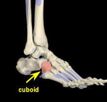
The cuboid bone is located on the outside of the foot. In front, it articulates with the metatarsal bone through the tarsometatarsal joint. Posteriorly, it articulates with the calcaneus through the calcaneocuboid joint. On the inner side of the cuboid, it articulates with the 3rd cuneiform bone.
Particularly, incomplete displacement of the cuboid bone can cause swelling and pain on the outside of the foot. This is called cuboid syndrome which requires treatment by a doctor.
3. Cuneiform bones
The cuneiform bones are 3 small bones that make up the inside of the foot. They are divided into the 1st cuneiform (or medial cuneiform), 2nd cuneiform (or intermediate cuneiform) and the 3rd cuneiform (or lateral cuneiform).
The 1st cuneiform bone is the largest cuneiform and located on the inside of the foot. Posteriorly, it is attached to the navicular bone. In front, it is attached to the base of the 1st and 2nd metatarsal bones. On the lateral side, it is attached to the second cuneiform bone.
The 2nd cuneiform bone lies in between the 1st and 3rd cuneiform bones. It is flanked in front by the 2nd metatarsal bone and posteriorly by the navicular bone.
The 3rd cuneiform lies behind the 3rd metatarsal bone and in front of the navicular bone. At the lateral side of the 3rd cuneiform is the cuboid bone
Forefoot Bones Anatomy
The forefoot bones are located after the midfoot. They consist of 5 metatarsal and 14 phalanges bones. So, they are 19 in number.
1. Metatarsal Bones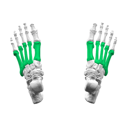
Like already mentioned, there are 5 metatarsal bones in the forefoot and the primarily help to connect the tarsal bones to the toes. They are numbered from inside to the outside as 1st, 2nd, 3rd, 4th, and 5th metatarsal bone.
The strongest, shortest metatarsal bone is the 1st metatarsal bone which transmits thrust in propulsion. It is the innermost metatarsal bone.
The 1st metatarsal bone is attached posteriorly to the 1st and 2nd cuneiform bones. The 2nd metatarsal is attached to all 3 cuneiform bones posteriorly. In a similar way, the 3rd metatarsal bone is attached posteriorly to the 3 cuneiform bone while the 4th metatarsal bone attached to the 3rd cuneiform and cuboid. The 5th metatarsal bone is attached to the cuboid bone posteriorly.
In front, all metatarsal bones are connected to the phalanges through the metatarsophalangeal joints.
The metatarsal bones are easily prone to stress fractures that happens when the foot is continually put under pressure. This is common in athletes especially basketballers and footballers.
2. Phalanges
The phalanges are small bones that make up your toes. They are 14 in number. Each toe has 3 phalanges (proximal, middle, and distal), except the big toe that consist of 2 phalanges (proximal and distal).
Between the proximal phalanges and the metatarsal bone is the metatarsophalangeal joints. In front of the proximal phalange is the middle phalange that is connected through the proximal interphalangeal joint.
In front of the middle phalange is the distal phalange that connected through the distal interphalangeal joint. The big toe houses 2 phalanges, the proximal and distal phalanges. The big toes do not have a middle phalange.